Saturday, March 25, 2017

Different Cages and Lumbar Lordosis

Week 6 & 7

Hi all, 

This week, I have been working a lot on my research paper and reading more articles related to spinal fusion surgeries and lumbar lordosis. A week ago, I found a study from The National Center for Biotechnology Information called Improvement of Segmental Lordosis in Transforaminal Lumbar Interbody Fusion: A Comparison of Two Techniques. Since the results of this study were so interesting, I ended up sharing this article with Dr. Larson and it eventually shifted the design of our own research project.

Review of the Study

The aim of this research was to analyze the effects of a traditional transforaminal lumbar interbody fusion (TLIF) approach versus a cantilever TLIF (c-TLIF) approach on segmental lordosis, disk height, and segmental coronal alignment. (All I really care about is the segmental lordosis data.) The investigators conducted a retrospective review on a total of 101 patients who received a TLIF between the years of 2006 to 2011 by three different spine surgeons. Of the 101 patients, 40 received the c-TLIF approach with a kidney-shaped disk cage and 61 received the traditional TLIF approach with a straight disk cage. With the c-TLIF approach, the surgeon inserted the cantilever cage in the disk space behind the anterior longitudinal ligament (ALL), so the disk would be situated as far forward as possible. With the TLIF approach, the disk cage was also placed in the anterior region of the disk space but in a slanted direction. Neither of the disks had any inherent lordosis, meaning that the design of the graft would not curve the spine once placed in the space.


Image of the cantilever TLIF cage and TLIF cage
Rice, James W. "Fig. 1" The National Center for Biotechnology Information, Cara L. Sedney, Scott D. Daffner, Justin W. Arner, Sanford E. Emery, John C. France, vol. 6, no. 3, 2016, p. 230, The National Center for Biotechnology Information,2016, West Virginia. The Global Spine Journal, https://www.ncbi.nlm.nih.gov/pmc/articles/PMC4836934/


Image of the Anterior Longitudinal Ligament

Augusta University. "Anterior Longitudinal Ligament." Augusta University Health, http://www.augustahealth.org/spine/spine-glossary/spine-glossary-anterior-longitudinal-ligament-(all).

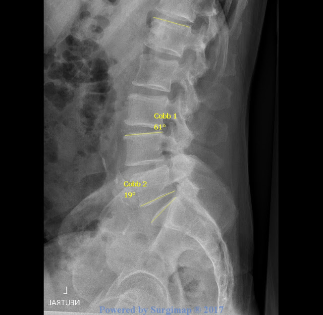
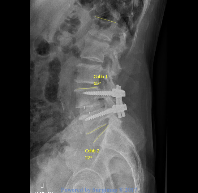
The investigators of the study used the Cobb method to take disk height, segmental coronal alignment, and segmental lordosis measurements from x-rays. (Again, segmental lordosis refers to the vertebrae of the spine where the spinal fusion was performed.) The investigators of the study took the measurements at the following times:


t0: x-rays taken on the day of surgery
t1: x-rays taken at patient's first postoperative follow-up (the average time for both the c-TLIF and TLIF group was six weeks)
t2: one year follow-up
t3: final follow-up (c-TLIF group: average final follow-up time was 26 months; TLIF group: average final follow-up time was 22 months)

The researchers subtracted preoperative measurements from every other following measurement. The mean change for each measurement was computed for both the c-TLIF and TLIF groups. Finally, statistical analysis was performed to compare the two groups' data by using a two-tailed t-test. 

Results 

c-TLIF Group
Mean ΔSegmental Lordosis (SL) from t0, t1, t2, t3 [°]
Mean Preoperative SL Value (°)
t0
t1 (mean= 6 weeks first follow-up)
t2
T3 (mean= 26 months final follow-up)
3.1
7.8
6.5
5.5
4.9


TLIF Group
Mean ΔSegmental Lordosis (SL) from t0, t1, t2, t3 [°]

Mean Preoperative SL Value (°)
t0
t1 (mean= 6 weeks first follow-up)
t2
t3
(mean=22 months)
5.6
4.0
1.8
0.4
0.1

After statistical analysis, the investigators found that patients in included in the c-TLIF category had segmental lordosis improved significantly (p<0.0001) compared to patients who received the straight TLIF cage.

So how does this relate to my research? Well, I sort of indicated in my last blog post that I would be looking at different cage types as a result of learning the implications of this Rice study. My advisor, Dr. Larson, believes that companies who specialize in spine instrumentation will be very interested to see if the type of cage used in a MIS TLIF affects lordosis. Yet, this study analyzes the effects of a traditional TLIF on segmental lordosis whereas mine will analyze the effects of a minimally invasive TLIF on both global and segmental lordosis. My research has never been done before, so the results of my research will be of great significance to spine surgeons since the preservation and restoration of lordosis is the optimal goal of many spine surgeries.

I intend to imitate this Rice study for my own research project by dividing my research subjects into groups based on the cage type used in the surgical procedure, compare patients' delta preoperative vs. postoperative measurements, and conduct a two-tailed t test to contrast the two groups' data.

That is all for this week! This week's yoga pose is Child's Pose! Click the link and try it out.

Until next time,

Hannah

Tuesday, March 14, 2017

March Madness: What I have been up to these past two weeks

Week 4 & 5


Greetings from snowy Coeur d'Alene, ID!

I am so sorry for my late blog post! I have been traveling a lot these past two weeks and working at my internship, so it's been hard to write this post but here I am! This week I started my internship at CDA Spine and so far it has been such a cool experience shadowing Dr. Larson, working with x-ray technicians, and gathering the data for my research project.

On Monday, I learned how to use the Surgimap software, which is a computer program that allows researchers to take spinal parameter measurements from patient x-rays. Surgimap is an extremely helpful tool because it allows doctors and researchers to easily find a patient's Sacral Slope, Pelvic Incidence, Pelvic Tilt, Lumbar Lordosis, Thoracic Kyphosis, Sagittal Vertebral Axis, and Cobb Angle! Without this software, these parameters would be much more complicated to take. Moreover, the aforementioned measurements above are crucial to understanding the health of a patient's spine, especially if the patient is undergoing surgery. These angles allow the surgeon to make decisions on how to better correct or mend the spine to a desirable state for the patient's pathology.

To measure lordosis using the Surgimap software, you can use the Cobb measuring instrument or the Lumbar Lordosis measuring instrument. The only difference between the two is that the Cobb measurement will create a positive value and the Lumbar Lordosis measurement will create a negative value. I used the Cobb method because that was the way I was taught!

Here is a picture of the Surgimap program:


(You can't really see the instrument options in the tool bar, but I'd use the very first option, >Cobb. For some reason this picture doesn't show the pelvic incidence tool.)

For the purposes of my research, I needed to measure my research subjects' preoperative, post operative, and most recent global lordosis (L1-S1) and preoperative and postoperative segmental lordosis (the level where they received their one-level fusion). For example, if a patient received a MIS TLIF to remove a degenerative disc between the L4-L5 vertebrae, I would measure the preoperative to postoperative change in lordosis for the L4-L5 vertebral segments. In addition to measuring lordosis, I would measure a patient's pelvic incidence and pelvic tilt if both of the hips were visible in the x-ray. However, it was really rare to find a preoperative and postoperative x-ray with both hips. Even if a patient had a preoperative and postoperative x-ray with both hips the pelvic incidence, which is supposed to be a constant value for each patient, would change! The x-ray technician that I worked with said that even if you align both of the circles with the hips  and the straight line with the endplates of the S1 vertebra like you did in the first x-ray (the preoperative x-ray), you can still get different measurements! Measuring pelvic incidence and pelvic tilt were by far the most challenging for this reason; also, it was hard to tell if the circles in the x-ray were gas bubbles or actually the hips! (I'll post some pictures later in this post to show you what I am talking about.) There was one time where I measured a pre-op and post-op pelvic incidence and the values came out to be the same. I was very proud!

Image of Global Lordosis (L1-S1)


Initially, I found the program to be complex because I was not familiar with reading x-rays and understanding where to draw the lines along the endplates — sometimes the imaging of the x-ray made it even more difficult to find the endplates of the vertebra. There were times where I would just have to make my best guess! The x-ray technician I was working with advised me to be consistent with the method I chose to use because there are a variety of ways to take these measurements. (It is all very subjective and arbitrary.) Eventually, I gained confidence in my method. I decided to draw the lines from the front endplate to the back endplate of the vertebra.

Taking this data was time consuming! It took me three six-hour work days to get measurements for 95 patients!

Here are some pictures I took of my data to give you a better idea! (FYI, patient names were removed for confidentiality reasons, but I will randomly make-up a name for the purposes of explaining...and because it’s fun!)


Extra: Here is a picture of an x-ray I measured with pelvic incidence and pelvic tilt


This here is Bob's preoperative x-ray. The segment where the surgery will be performed is the L4-L5 segment (indicated by Cobb 2). His global lordosis is 61 degrees and his segmental lordosis is 19 degrees. Remember, the aim of my research is to see what effects (if any) MIS TLIF has on global and segmental lordosis.

Bob L4-L5 Lumbar Fusion: Preoperative Image


This is Bob's six-week postoperative x-ray. His global lordosis increased 5 degrees and his segmental increased 3 degrees. While Bob’s global and segmental lordosis increased, we must remember that he is one subject and that his "trend" is not applicable to my larger population — at least not yet. When I was taking these measurements, it was hard to generalize a trend with pre-op and post-op change, which is why I will have to perform a two-tailed t-test to test the significance of the pre-op to post-op lordotic change.

Bob L4-L5 Lumbar Fusion: Postoperative Image 

The three white lines in the disc space illustrate a traditional TLIF cage. There are two different cage types in my study: a TLIF cage and a PLIF cage. The TLIF cage is shaped like a pear, made from polyetheretherketone (PEEK), and has five degrees of inherent lordosis. In my study, TLIF cages were more prevalent in patients who received a MIS TLIF from 2012 to 2014. Bob must have been a patient during that time period because he received a TLIF cage. The PLIF cage that Dr. Larson uses looks like an elongated oval and is made of Tritanium. The PLIF cage has 6 degrees of inherent lordosis. (In my next blog post, I will further elaborate on the different cage types and their relevance to my study.)

Here is a picture of an x-ray with the PLIF Cage:


Here are two pictures of the sample PLIF cage used in my study: (the sample is MUCH bigger than the actual cage used in surgery.)


In addition to taking measurements, I got to shadow Dr. Larson around in clinic. Before Dr. Larson and I made our way to the exam rooms, Dr. Larson would show me the patient’s MRI and where the lumbar fusion was performed on the spine. Most of the patients I saw had already received surgery and were there for a post-op or one year check-up. For example, the first patient I observed had a L4-L5 fusion back in the fall and, for the most part, seemed to be in good spirits. Patient 1 complained about leg tingling and numbness in the upper thigh, so Dr. Larson checked the patient’s reflexes to see what nerve was hurting. Dr. Larson would ask the patient to move into certain positions that are shown below.


Flexing the foot upward was the hardest movement for Patient 1 to perform so the L4-L5 nerves were of concern. Patient 1’s MRI looked perfect in Dr. Larson’s words, so Dr. Larson wasn’t concerned that the nerve was pinched. To be sure, a CT scan was ordered. I found Patient 1’s scenario to be representative of the other patients' I observed. The majority of the patients were mostly in pain, but Dr. Larson explains that the pain is a result of an “insulted” nerve after surgery. Like Patient 1, CT scans were order for almost all the patients for reassurance.

Aside from going to clinic, I was able to watch an x-ray, an MRI, and a Kyphoplasty. A Kyphoplasty is a surgery used to treat compression fractures in the spine. People with osteoporosis are more likely to need this procedure. Dr. Larson uses a minimally invasive technique when he performs a Kyphoplasty. At the end of the Kyphoplasty I watched,  Dr. Larson shaped the cement into a stick-figure man! Click Here to watch an animated video that illustrates the procedure.


Well, that is all for this week. After shadowing Dr. Larson in clinic, I realized how important it is to take care of your spine! Dr. Larson says twisting is one of the best things you can do to decompress the spine. Hold the following yoga pose for five minutes on each side! (Watch the video)

Half Lord of the Fishes Pose 


Ciao,

Hannah   

Wednesday, March 1, 2017

It's All in the Details: The Specifics of an Interbody Fusion

Week 3

In my very first blog post, I introduced that I will be analyzing Minimally Invasive Spine Transforaminal Lumbar Interbody Fusion (MIS TLIF) for my Senior Research/AP Research Project. So what is exactly is a lumbar fusion?

The basic definition of a lumbar fusion surgery is a procedure that joins or fuses two or more vertebrae. Spinal fusion surgeries are used to treat or help alleviate pain caused by a variety of spinal pathologies like Degenerative Disc Disease, Spondylolisthesis, Neurogenic Claudication, and more. There are multiple ways a lumbar fusion can be performed, but the approach ultimately depends on the patient’s condition and area of pain. For example, an Anterior Lumbar Interbody Fusion (ALIF), a Posterior Lumbar Interbody Fusion (PLIF), an Extreme Lateral Interbody Fusion (XLIF), or a Minimally Invasive Transforaminal Lumbar Interbody Fusion (MIS TLIF) can be performed. Moreover, a spinal fusion isn't limited to the lumbar spine, vertebrae can be fused on cervical and thoracic spine as well. As aforementioned above, the location of the damaged disc (in conjunction with the symptoms) helps the surgeon decide where to make the incision/direct the surgery. An ALIF, PLIF, and XLIF all are surgeries used to replace a damaged intervertebral disc with a cage filled with bone graft. On the other hand, TLIF is a bit more complex.

For the specific purposes of my blog, we will be considering the contexts and implications of these surgeries within the lumbar spine as this is the part of the spine where lordosis occurs. In this next section, I provide short summaries of these procedures and how they differentiate from MIS TLIF.


Anterior Lumbar Interbody Fusion (ALIF)

ALIF is commonly performed on a very specific location on the lumbar spine, the L4-L5 vertebrae or L5-S1 portion. According to Spine Surgery: Techniques, Complication Avoidance, and Management, one of the texts books Dr. Nicol loaned me, an ALIF procedure "allows for restoration, or at least improvement, of normal lumbar lordosis." In addition, ALIF is a more favorable procedure compared to PLIF because there is a lower complication rate. I won't go into too much specifics about ALIF, but I included this video because it demonstrates the wide variety of spinal procedures and techniques.

Click here to watch the video that describes an ALIF.

Posterior Lumbar Interbody Fusion (PLIF)

PLIF is very similar to ALIF. Instead of performing the surgery from the ventral side of the body, it is performed from the dorsal side. (It's pretty intuitive.) According to Spine Surgery: Techniques, Complication Avoidance, and Management, PLIF gained popularity in the mid-twentieth century due to its creator, neurosurgeon Ralph Cloward. Initially, PLIF resulted in higher fusion rates, but more complications like blood loss, dural injury, and more. With time, better instrumentation allowed the surgery to become more successful. However, with PLIF, the thecal sac and nerves have to be pulled aside to get to the disc, amplifying the likelihood for damaging the nerve roots or the dura mater.

Click here to watch an animated PLIF.

Extreme Lateral Interbody Fusion (XLIF)

XLIF is very similar to the aforementioned spinal fusions above. Unlike ALIF or PLIF, XLIF cannot be performed on L5-S1 or L4-L5 (in some cases) . Moreover, the surgery is performed from the side of the body.

Click here to watch XLIF.

Transforaminal Lumbar Interbody Fusion (TLIF)

A traditional TLIF is performed posteriorly. This approach fuses the spine together with pedicle screws and rods to help fuse the spine together. A bone graft is inserted between the vertebrae and along the back the vertebrae because parts of the vertebrae have to be removed in order to perform surgery on the damaged discs. Eventually the bone graft will create a "bone bridge" between the damaged vertebrae.

Click here to watch TLIF.


Minimally Invasive Spine Surgery

Minimally Invasive Spine surgery is a procedure that uses smaller incisions than traditional open back surgery. This approach decreases damage to muscles and ligaments around the spine by utilizing equipment like endoscopes, microscopes, and retractors. Moreover, a minimally invasive approach is associated with less blood loss, faster recoveries, and increased patient satisfaction.

Here is a short video introducing the MIS technique featuring Dr. Jeffrey J. Larson, M.D., my external advisor:


Here is a MIS TLIF Video also featuring Dr. Larson:


I hope that you found this blog post helpful!

Until next time,

Hannah


P.S. this weeks yoga pose is extended puppy pose.


Sunday, February 26, 2017

Let's Break it Down

Week 2


Hi all, 

It's all about the basics, right? Here is some background information for you guys to help you better understand my research.

The Spine Anatomy 

The spine can be broken down to into three parts—the cervical spine, the thoracic spine, and the lumbar spine—all of which are comprised of vertebrae. The cervical spine has seven vertebrae, the thoracic spine has twelve vertebrae, and the lumbar spine has five vertebrae. Below the lumbar spine is the sacrum, which has five vertebrae that are joined together. The purpose of the sacrum is to connect the lower spine to the pelvis and sacroiliac joints Lastly, below the sacral spine is the coccyx, which is also known as the tailbone. Click here to view a really cool video that illustrates the anatomy of the spine.


In between the vertebrae are intervertebral discs, structures that cushion, support, and allow the spine to move. The disc anatomy can be broken down into two parts: the annulus fibrosis and the nucleus pulposus. The annulus fibrosis, which is the outer part of the disc, is made of strong collagen fibers. If you use your latin, can you guess what the nucleus pulposus refers to? Just like it sounds, the nucleus pulposus refers to the gelatinous center of the disc, which is comprised of a loose network of fibers. 

A healthy disc is a disc that is hydrated. When humans are born, the fluid inside the intervertebral discs is 80% water. As humans age, the discs are unable to hold as much fluid as they used to. As a result, the disc loses its robust shape and compresses, shortening the spine. This is why humans shrink as they age. 

Remember Degenerative Disc Disease? For a reminder here is a video that illustrates the specifics: 


Lordosis, Kyphosis, and Sagittal Balance

A healthy spine has two curves: kyphosis, the outward curve of the spine, and lordosis, the inward curve of the spine. Both the kyphotic and lordotic curves serve to balance one another and this is known as sagittal balance. A healthy spine has normal lordosis and normal kyphosis. According to a study called Sagittal Plane deformity: an overview of interpretation and management published by The National Center for Biotechnology Information (NCBI), there is a healthy relationship between the kyphotic and lordotic curves of the spine. However, if one curve is pronounced more than the other curve, this creates sagittal imbalance, sometimes other spinal deformities, and back pain. An over-pronounced lordotic curve is known as swayback and an over-pronounced kyphotic curve is known as hunchback or hyperkyphosis. 

So how does this all relate to my research? Unfortunately, that is for next week's post. 

Don't forget to try out this week's yoga pose: Triangle. Comment down below and share your thoughts! 

Ciao, 

Hannah 


Shoulda, Coulda, Woulda

Week 1

Do you ever have those self-scrutinizing moments where you reflect and try to pin-point at what time in your life you messed up? If so, yay I am glad I am not alone. If you need more "inspiration", I highly recommend conducting research as a seventeen year-old. Because the material is so unfamiliar and difficult, research really makes you analyze everything...including your life choices. So the other day, I figured out where I went wrong in my life. I didn't go to medical school.

Since this blog is supposed to explain my research experiences, I thought I'd take the time to chronicle my newest challenge: understanding medical literature. This was me when I first started reading articles for my research. Yes. I was a bit overwhelmed.

WHY DO I NOT UNDERSTAND??! I SHOULD HAVE GONE TO MED SCHOOL!!


When the literature is peppered with words like "osteotomies", "percutaneous screw fixation", and "cauda equina compression", how am I supposed to react? More importantly, how am I going to be able to write a five thousand word paper about all of this material I currently don't understand? Key-word currently. In another less scrutinizing self-reflecting moment, I realized that while I currently don't understand the material, it doesn't mean that I won't be able to eventually (even though it feels like I never will). 

I realized that in order to understand more complex articles that are related to my research, I needed to have a basic understanding of the spine anatomy. So like a true BASIS student, I went looking for resources. Fortunately, the Flagstaff medical community is so supportive and willing to help high school students like me. I reached out to a local neurosurgeon, Dr. Bradley Nicol, M.D., and he loaned me two of his medical school text books! So over the next few weeks, these guys will be my best friends!


Receiving help has restored my optimism and enthusiasm for this new, challenging adventure.  I actually really enjoy perusing the text books and learning about the spine. I feel like an actual medical student, which makes me feel super cool! I am excited that I have been given this opportunity as a senior in high school to explore and apply my skills to a foreign field of study.

This week's yoga pose: Sphinx Pose. According to the Yoga Journal, this pose helps mitigate stress and strengthen the spine. Click here for step-by-step instructions on how to do the pose. Comment down below if you tried this at home!


See you next week,

Hannah

P.S.  I am sorry for the pedestrian moral!


Thursday, February 23, 2017

Introduction


What do school, car rides, studying, cuddling with your dog, and watching The Bachelor all have in common?

Did you say nothing, Mr. Lamb? If so, you’re wrong.
The correct answer is… sitting!

Like most of you, I spend the majority of my days sitting. I sit when I go to school, I sit on the way to and from school, I sit when I eat, I sit when study, and even when I workout whether it be sit-ups or spin class, I sit. In summation, I sit a lot and you all sit a lot too! So why is this bad?

It is kind of obvious but sitting compresses the discs in the spine, creating pain and discomfort. (We’ve all been there.) Twisting in your chair and circling your neck are good solutions to help relieve the pain in the short-term, but unfortunately these exercises won’t be enough to maintain a healthy spine in the long run. Over time, the discs that cushion the vertebrae in your spine will  (most likely) degenerate. This condition is known as Degenerative Disc Disease. The compressed disc causes persistent nerve pain otherwise known as radiculopathy. Sometimes this nerve pain can radiate down your arms and legs and this is called radiculitis. The moral of the story is that sitting has bad consequences if you don’t modify your lifestyle. In some cases this disease is associated with the normal process of aging. Either way, you are at risk!

Degenerative Disc Disease cannot be treated, but radiculopathy and radiculitis can be treated by a Minimally Invasive Transforaminal Lumbar Interbody Fusion (MIS TLIF). While MIS TLIF is known to have many short-term, postoperative benefits, the long-term effects of MIS TLIF, specifically on lordosis, are largely unknown among the neurological community. (Don’t worry. I will be writing another blog post delineating what all this medical jargon means and why the preservation of lordosis is important for long-term spine health.) My research will be looking to see if MIS TLIF has a positive, negative, or neutral effect on lordosis by comparing patient preoperative and postoperative spinal parameter measurements.

My external advisor for my project is Dr. Jeffrey J. Larson, M.D., a neurosurgeon in Coeur d’Alene, Idaho, and my internal advisor is my fabulous Statistics teacher, Chris Lamb. I will be interning at CDA Spine in March. Click here to visit the website.

Lastly, I hope to incorporate some Yoga poses that will help relieve your spine into my weekly posts. Might as well be proactive right?

No matter how boring my research may sound, it applies to all of you! So, I suggest you join me on my journey and pay close attention:)

Until next time,

Hannah